
A small home for a big microscope
Dr. Christoph Diebolder wants to know what holds the world together at its innermost core. He wants to understand the biochemical processes by which the smallest pieces of the puzzle of life fit together, and to render visible what happens when molecules within a cell collide.
We put a lot of effort into making sure our microscopes are happy.
Since February 2020, the structural biologist has been director of the newly established Core Facility for Cryo-Electron Microscopy (cryo-EM) run by the Charité – Universitätsmedizin Berlin together with the Max Delbrück Center for Molecular Medicine in the Helmholtz Association (MDC) and the Leibnitz-Forschungsinstitut für Molekulare Pharmakologie (FMP). Previously, he spent more than ten years in the Netherlands, where he worked as a scientist at the Netherlands Centre for Electron Nanoscopy (NeCEN), among others. Located on the Buch campus, the cryo-EM facility has been in test operation for almost a year, and officially went into regular operation in March. With cryo-EM, it is possible to render nanometer-sized biological molecules visible “at a resolution that rivals crystallography,” says Diebolder enthusiastically. He will give his welcome lecture on April 13.
Cryo-EM has a decisive advantage over X-ray crystallography, explains Professor Oliver Daumke, as it can also be used to image three-dimensional structures such as proteins. Daumke’s research at the MDC focuses on proteins that carry out important functions within the cell by deforming cellular membranes during energy consumption. “While crystallography produces an image of an isolated protein, the special thing about cryo-electron microscopy is that it allows us to look at proteins not only in isolation, but also in their cellular environment,” says Daumke. “And it eliminates the need to crystallize them beforehand.”
Cryo-microscope provides highest quality data

Sensitive giant: The four-meter-high cryo-transmission electron microscope cannot tolerate vibrations from passing cars or temperature fluctuations.
The centerpiece of this technology platform is a four-meter-tall cryo-transmission electron microscope (cryo-TEM). It stands like a huge vault in a high-ceilinged white room, where a ventilation system emits a constant hiss. This is a necessary feature, as the device is just as sensitive as it is large, and unable to tolerate temperature fluctuations or excessive humidity. The room’s humidity is therefore kept under 20 percent, which means visitors get thirsty very quickly. Vibrations and electromagnetic fields are also problematic. A concrete foundation 1.25 meters thick has therefore been placed underneath the building to cancel out underground vibrations. Otherwise, the campus bus passing by 100 meters away would interfere with the measurements. And with its double walls, the building is also a sort of house within a house. “We put a lot of effort into making sure our microscopes are happy,” says Diebolder.
Diebolder clearly remembers the moment he switched on the cryo-TEM for the first time a year ago: “This can be a moment of great disappointment. But in this case it was wonderful, because we received very high-quality data.” Researchers in Berlin can now use the state-of-the-art technology for their own projects, with the support of the cryo-EM team. This support can include preparing samples, taking the measurements, and assisting in the evaluation and modeling of the data. Alongside Diebolder, Dr. Thiemo Sprink (MDC) and Metaxia Stavroulaki (Charité, funded by the neighboring Leibniz-Forschungsinstitut für Molekulare Pharmakologie) work in the new facility – which is a plain, cube-shaped building clad in dark-brown wood. A desk runs lengthwise through the room with four workstations, each equipped with several screens. The view from the window is currently 2,400 m2 of sand, which will soon be the site of another construction project: This is where the MDC’s Optical Imaging Center (OIC) is scheduled to open in one year’s time.
Flash-frozen preparations in their natural structures
Diebolder became passionate about electron microscopy while studying technical biology in Stuttgart. That was in the 2000s, and cryo-EM did not exist there at the time. Nevertheless, Diebolder became fascinated, as even a conventional electron microscope enables a view of individual atoms. This is possible because electron beams achieve a much higher resolution than light, thanks to their shorter wavelength. However, the samples must first be dehydrated, chemically fixed and stained with heavy metals to enhance image contrast. In other words, what the electron beam makes visible is not the natural state of a molecule, but what remains of it after this treatment.

The cryo-EM team (from left to right): Dr. Christoph Diebolder, Dr. Thiemo Sprink and Metaxia Stavroulaki in front of the four-meter-high cryo-electron microscope.

The newly constructed building for the Cryo-EM Core Facility is located as far away from busy roads as it is possible to get on the Campus Buch.


It is only thanks to cryo-EM that scientists no longer have to interpret artifacts, but can actually look at natural structures. In 2017, the Swiss Jacques Dubochet, the German-American Joachim Frank and the Scot Richard Henderson were awarded the Nobel Prize in Chemistry for developing this new type of microscopy. In this process, molecules are cooled so quickly that ice crystals cannot form. Instead, glasslike amorphous ice is formed, which tightly encloses the molecules in their natural state. The electron beam can penetrate the ice layer. A camera installed in the lower part of the microscope records the image. A computer calculates an exact three-dimensional image of the molecule from what are often hundreds of thousands of photos – with unprecedented accuracy and detail. The resulting images often reveal an unexpected beauty and symmetry that an architect could not have designed better.
300 nanometers are the maximum
The remote realms into which the researchers are penetrating with the electron beam can also be seen in the contrast between the size of the microscope and the small size of the sample it can illuminate. A cell one micrometer in diameter would be too thick; 300 nanometers are the maximum. To be able to prepare such delicate specimens, the researchers at the facility have another top-class instrument at their disposal: a dual-beam FIB SEM, which is a cryo-scanning electron microscope with a focused ion beam. An electron-permeable lamella is milled out of the flash-frozen sample, such as a human cell, with the ion beam. To get precisely the section to be viewed in the cryo-TEM, Metaxia Stavroulaki can mark the corresponding molecules beforehand using a cryo-CLEM, a correlative light electron microscope. The lamella, this very tiny sample, then goes into the cryo-TEM.
In addition to providing services to researchers and their projects, Diebolder and his colleagues hope to further develop and refine this work flow – from cryo-CLEM to dual-beam FIB SEM and cryo-TEM to data preparation and model calculation. “We would like our work to advance structural biology in a methodical way,” says Diebolder, describing his vision. “Our goal is to move from in vitro to in situ, that is, to observe processes directly in the cell.”
The ribosome as a universe
In parallel, the cryo-EM team is working on its own projects. Thiemo Sprink has devoted himself to bacterial ribosome – or more precisely, one of the atoms that make up a ribosome. There are hundreds of thousands of them. He hopes to find out what this one atom is for. His search began in 2015 as part of his doctoral thesis. He has calculated a three-dimensional ribosome model from the images of hundreds of thousands of ribosomes.
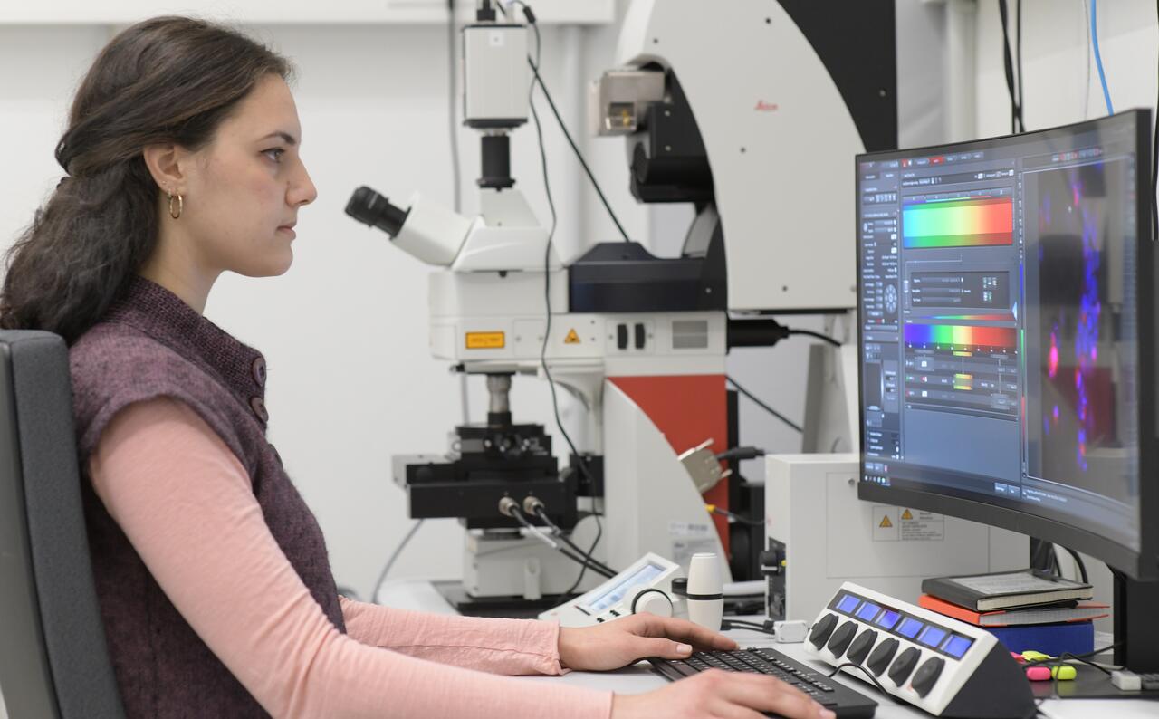
Something that looks like a gray sponge appears on the computer screen. With a click, Sprink can change the representation. The sponge turns into a crumpled grid. Based on the density of the lines, Sprink can identify the building blocks that make up the ribosome. The density also shows where molecules interact with one another or how and where a single ion steers amino acid side chains. He marks RNA either green or yellow, proteins blue or red – creating a tangle of colored, intertwined bands. “Simply put, you could say that where the colors mix, interactions between molecules are probably taking place,” Sprink explains. He can then take a closer look at these spots by conducting further experiments under the microscope. He clicks here and there, switches back and forth between the views, dipping into the universe of the ribosome. With the help of VR goggles, he can even immerse himself completely, walking through the ribosome as if it were a labyrinth.
“It’s truly exciting,” Diebolder says. “Sometimes I’d like to be less of a manager and more of a scientist.” Still, he couldn’t have imagined anything better than getting the Charité facility up and running. His logistical skills will also be in demand when the MDC’s Optical Imaging Center opens, for at that point some of the equipment from his facility will move to the new building just across the street. Close collaboration with MDC scientists is planned – including with the new junior research group “In Situ Structural Biology” headed by Dr. Misha Kudryashev, who will start work in the summer.
Text: Jana Ehrhardt-Joswig
Further information


- Buch campus to get ultralow temperature microscopes
- Institute of Medical Physics and Biophysics at Charité
- Press release by FU Berlin: German Research Foundation approves funding for new high performance microscopes (German only)


